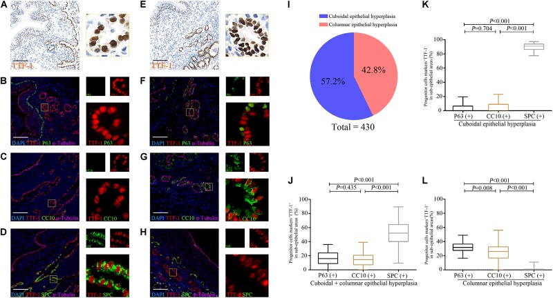
FIGURE 5.
Expression of TTF-1, P63, CC10 and SPC protein in the sub-epithelium areas of the dilated bronchioles in bronchiectasis. Two major abnormal patterns are observed in the sub-epithelium areas of control and dilated bronchioles from patients with bronchiectasis with TTF-1 staining: cuboidal epithelial hyperplasia (A–D) and columnar epithelial hyperplasia (E–H). We confirm that most progenitor markers (P63, CC10 or SPC) (green) co-localize with TTF-1 (red) (B–D,F–H). We count 430 areas (10 areas per patient) with epithelial hyperplasia. 57.2% (n = 246) of areas present with cuboidal epithelial hyperplasia, whereas 42.8% (n = 184) of areas yield columnar epithelial hyperplasia (I). The percentage of P63+TTF-1+, CC10+TTF-1+ and SPC+TTF-1+ are assessed in both cuboidal and columnar epithelial hyperplasia (J), only for cuboidal epithelial hyperplasia (K) and only for columnar epithelial hyperplasia (L), respectively. α-tubulin (pink) is stained as a ciliary marker for epithelium which is helpful to distinguish the sub-epithelium areas. Scale bar = 100 μm. CC10: club cell 10 kDa protein; SPC: surfactant protein C; TTF-1: thyroid transcription factor-1.

