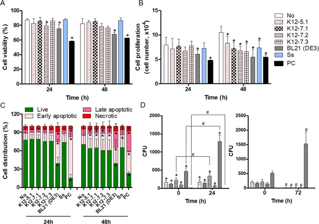
Figure 4.
Invasion of HOK-16B cells and induction of apoptosis by oral E. coli strains. HOK-16B cells were infected with oral E. coli strains and S. salivarius (Ss) at an MOI of 1000. As a positive control (PC), cells were treated with 0.5 µg/ml staurosporine. (A,B) Cell viability and proliferation were measured by trypan blue enumeration. *p < 0.05 compared to untreated cells by t-test. (C) The apoptotic stage was measured by flow cytometry using Annexin V-PI staining. *p < 0.05 compared to untreated cells by t-test. (D) HOK-16B cells were infected with bacteria at an MOI of 1000 for 2 hours, and the infected cells were treated with gentamicin for 1 hour. The cells were then lysed immediately or further incubated for 24 hours in antibiotic-free medium. The cell lysates were cultured on LB agar plates. *p < 0.05 compared to Ss by Mann-Whitney U test, #p < 0.05 by 2-tailed Wilcoxon signed-rank test. All graphs present the mean and standard deviation of two experiments performed in triplicate.

