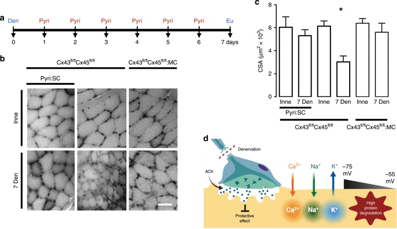
Fig. 3. Acetylcholine analog prevents cellular alterations in vitro, and increasing its half-life prevents the decrease of myofiber size in vivo.
Unilateral denervation of the sciatic nerve in Cx43fl/flCx45fl/fl:Myo-Cre (Cx43fl/flCx45fl/fl:MC) mice were performed. a Experimental design of in vivo acetylcholinesterase blockade. Den: denervation, Pyiri: pyiridostigmine, Eu: euthanasia. b Hematoxylin:eosin-stained cross-section of the FDB at day 7 post denervation. Inne: Innervated. Den: Denervated. c Cross-sectional area (CSA). N = 5; each value is the mean ± SEM. *p < 0.05 for Den compared with Inne muscles by Student’s t test. d Proposed model. Denervation (Den) eliminates the protective effect of ACh and leads to a reduction in acetylcholine (ACh) release resulting in an increase in Ca2+ and Na+ influx via de novo expressed non-selective membrane channels, increasing cytoplasmic concentrations of these ions. Consequently, a reduction in the RMP from −75 to −55 mV occurs due in part by K+ efflux and Ca2+ and Na+ influx; the increase in intracellular Ca2+ signal promotes protein degradation.

