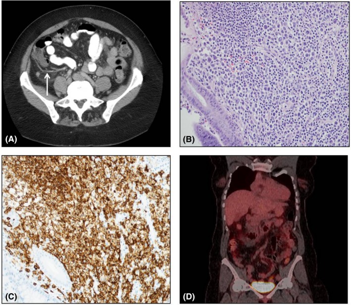
Figure 1.

A, CT scan showing dilated appendix with nonspecific periappendiceal inflammatory changes (white arrow). B, 200× H&E stain of appendix tissue revealing large atypical mononucleated cells. C. 200× CD20 stain of appendix tissue. D, PET‐CT at diagnosis revealing no evidence of hypermetabolism outside of the appendix
