Fig. 4.
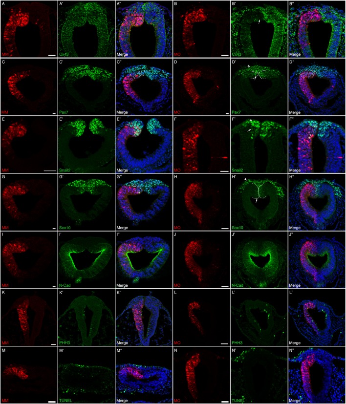
Cx43 depletion decreases the number of premigratory neural crest cells in the absence of any changes in cell proliferation or cell death. Representative transverse sections taken through the midbrain and hindbrain of HH8+ to HH9 embryos after unilateral electroporation of the neural tube at HH7+ to HH8− (2ss–3ss) with either the control morpholino (MM) or Cx43 MO and immunostaining for various neural crest cell markers. In MM-treated embryos (A–A″,C–C″,E–E″,G–G″,I–I″,K–K″,M–M″), there is no decrease in the level of Cx43 in the neural tube (A′) and no change in any of the premigratory neural crest cells as identified by Pax7 (C′), Snail2 (E′), Sox10 (G′). Similarly, there are no changes in N-cadherin (I–I″), a marker of neural tube cells, cell proliferation [pHH3 (K–K″)]; or cell death [TUNEL (M–M″)]. In Cx43 MO-treated embryos (B–B″,D–D″,F–F″,H–H″,J–J″,L–L″,N–N″), however, there is obvious knockdown of Cx43 in the neural tube (B′, arrow) and also a reduction in the premigratory neural crest cell population observed after Pax7 (D′, arrow), Snail2 (F′, arrow) and Sox10 (H′, arrow) immunostaining. Interestingly, N-cadherin (J–J″) is not affected. Cx43 MO-mediated loss of Cx43 also does not seem to alter levels of either cell proliferation (L–L″) or cell death (N–N″). Additionally, in HH9 embryos, a small number of MO-positive neural crest cells begin to emerge from the neural tube (D′,F′,H′, arrowheads; neural tube indicated by dashed lines). Scale bars: 20 µm (A–K″,M–N″); 50 µm (L–L″).

