Fig. 8.
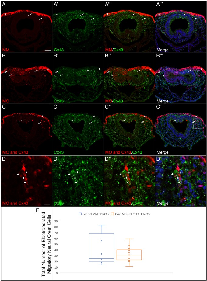
Co-electroporation of Cx43 MO and a full-length Cx43 construct refractory to the MO rescues the delayed neural crest cell emergence from the neural tube. Representative transverse sections taken through the midbrain and hindbrain of HH11 embryos after dorso-ventral electroporation at HH8 of the dorsal surface of the neural folds with the control MM (A–A‴), Cx43 MO (B–B‴), and co-electroporation of the dorsal neural folds with Cx43 MO on the dorsal surface and full-length rat Cx43 in the neural tube (C–D‴). Electroporation with the control MO does not alter levels of Cx43 in either the ectoderm (A″, asterisk) or the neural crest cells (A″, arrows) while there is a significant reduction in Cx43 in the ectoderm (B″, asterisk) and neural crest with the Cx43 MO (B″, arrows). Co-electroporation of the Cx43 MO and full-length rat Cx43 does not rescue the knockdown in the ectoderm (C″, asterisk), which does not receive the full-length rat Cx43, but is able to rescue the levels of Cx43 in both the neural tube and the migratory neural crest cells (C″, arrows). The box in C‴ is shown at a higher magnification in D–D″, and shows the rescue of the Cx43 MO-electroporated neural crest cells (arrowheads) to almost normal levels of Cx43. This restoration of Cx43 results in normal numbers of neural crest cells emerging from the neural tube (C″, arrows). (E) Data were quantified with an un-paired Mann–Whitney (Wilcoxon) test, which showed no significant difference in the number of migratory neural crest cells between control MM-treated embryos and Cx43 MO embryos co-electroporated with full-length Cx43 (P=0.89), graphically depicted by a box and whiskers plot as in Fig. 5. Scale bars: 50 µm (A–C‴), 10 µm (D–D‴).

