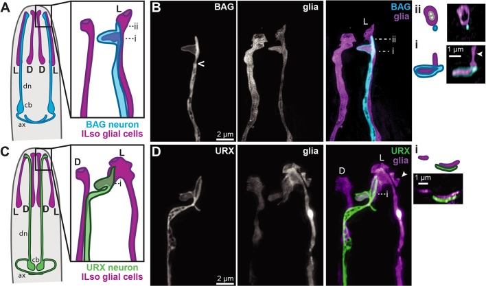
Fig. 1.
BAG and URX dendrites form specialized contacts with a single glial cell. (A,C) Schematics of a C. elegans head (nose is at the top) showing BAG (A, blue) or URX (C, green) relative to the inner labial socket glia (ILso, purple). Dorsal (D) and lateral (L) pairs of glia are indicated; an additional ventral pair is not shown. cb, neuronal cell bodies; dn, dendrites; ax, axons. (B,D) Single-wavelength and merged super-resolution images of BAG (blue, flp-17pro) or URX (green, flp-8pro) and ILso glia (purple, grl-18pro) acquired with structured illumination microscopy. Caret indicates the base of the cilium. Bi and Bii show cross-sectional views and schematics of the indicated image planes. (Bi) BAG wraps around a thumb-like protrusion of the glial cell. A second protrusion is visible in this view (arrowhead). (Bii) The ILso glial cell forms a pore through which two unlabeled neurons access the environment; BAG does not enter this pore. (D) The URX dendrite ‘jumps’ from the dorsal to lateral ILso and forms a sheet-like contact with a protrusion from this glial cell (cross-section in Di). A second protrusion from the glial cell, likely the BAG-associated thumb, is partly visible (arrowhead).

