
Fig. 2.
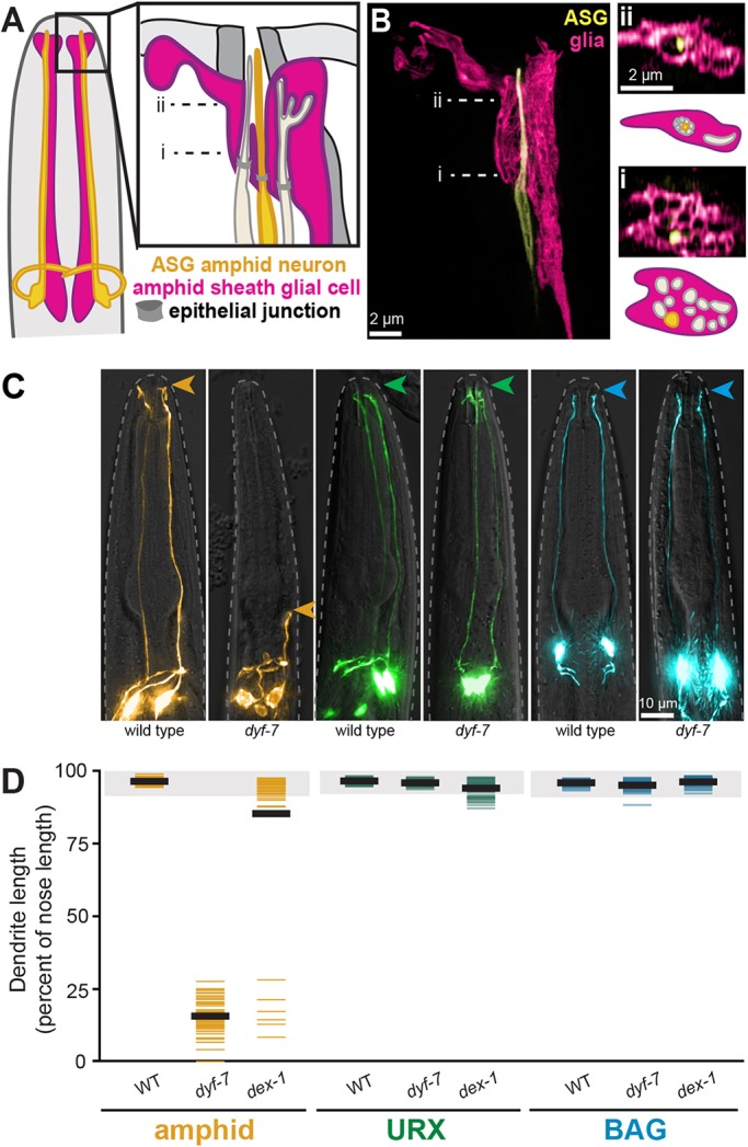
URX and BAG dendrites develop by a mechanism distinct from the amphid. (A) Schematic of a C. elegans head showing representative amphid neurons (ASG neuron, yellow; other neurons, tan) and the amphid sheath glial cell (pink). Most amphid dendrites enter individual channels in the glial cell that converge to a central pore; some terminate in pockets within the sheath. (B) Single-wavelength and merged super-resolution images of ASG (yellow, ops-1pro) and the amphid sheath (pink, F16F9.3pro). i and ii show cross-sectional views and schematics of the indicated image planes. (i) Twelve individual channels in the sheath, one for each amphid dendrite, were previously seen only by EM. ASG can be seen inside one of these channels. (ii) The channels converge distally to a central pore with ASG inside it. (C) Wild-type and dyf-7 animals expressing markers for an amphid neuron (AWC, yellow, odr-1pro), URX (green, flp-8pro) or BAG (blue, flp-17pro). Arrowheads indicate dendrite endings. (D) Dendrite lengths in wild-type, dyf-7 and dex-1 animals, expressed as a percentage of the distance from cell body to nose. Colored bars represent individual dendrites (n≥50 per genotype); black bars represent population averages; shaded region represents wild-type mean±5 s.d. for each neuron type, which we define as ‘full length’. All amphid dendrites are affected by dyf-7 and dex-1 (Heiman and Shaham, 2009).