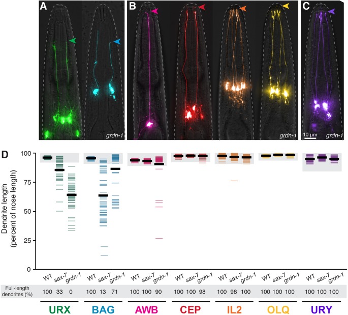
Fig. 4.
sax-7 and grdn-1 preferentially affect URX and BAG dendrite extension. (A,B) grdn-1 animals expressing fluorescent markers for (A) neurons that make specialized wrapping contacts with the ILso glia [URX (green, flp-8pro) and BAG (blue, flp-17pro)]; (B) neurons that enter glial pores [amphid (AWB, pink, str-1pro; two additional amphid neurons, AWA and AWC, are in Fig. S5), CEP (red, dat-1pro), IL2 (orange, klp-6pro) and OLQ (yellow, ocr-4pro)]; and (C) a neuron that does neither [URY (purple, tol-1pro)]. Arrowheads indicate dendrite endings. (D) Quantification of dendrite lengths in the indicated genotypes, expressed as a percentage of the distance from cell body to nose. Colored bars represent individual dendrites (n≥47 per genotype); black bars represent population averages. Shaded region represents wild-type mean±5 s.d. for each neuron type and the percentage of dendrites in this range (‘full-length dendrites’) is indicated below the plots. URX and BAG quantification is the same as in Figs 2 and 3.

