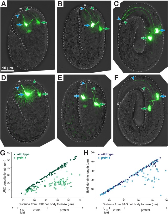
Fig. 5.
URX and BAG dendrite length defects arise during embryo elongation. (A-F) URX and BAG dendrite extension in (A-C) wild-type and (D-F) grdn-1 embryos at (A,D) 1.5- fold, (B,E) 2-fold and (C,F) pretzel stages. Embryonic URX and BAG were visualized using egl-13pro:GFP and distinguished by their cell body positions (URX cell body is more posterior, green arrow; BAG cell body, blue arrow). URX and BAG dendrite endings are marked with green and blue arrowheads, respectively. Asterisk indicates embryo nose. (G,H) Dendrite lengths plotted as a function of the distance from cell body to nose for (G) URX and (H) BAG in wild-type (filled circles) and grdn-1 (open circles) embryos. n≥70 per genotype.

