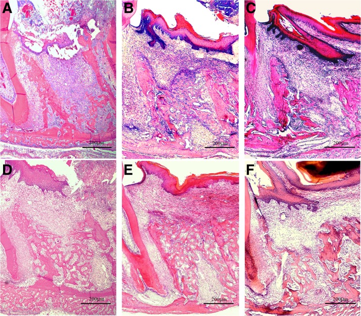
FIG. 5.
Histopathology on days 5 and 7 after tooth extraction. H&E staining; original magnification × 40. (A, D) Diode group; (B, E) CO2 group; (C, F) control group. Day 5 postextraction: new bone formation seen from the socket fundus in the diode group and the control group, with a lesser amount in the control group (A, C). In contrast, newly formed immature bone seen extending from the shallow to the middle layer of the extraction socket in the CO2 group (B). Day 7 postextraction: new bone formation seen from the fundus to the shallow layer of the extraction socket in the diode group and from the shallow to the middle layer with cross-linking pattern in the CO2 group. Denser cancellous bone is seen in the CO2 group than in the diode group (D, E). Cancellous bone formation in the extraction socket is immature, weak, and not continuous in the control group (F).

