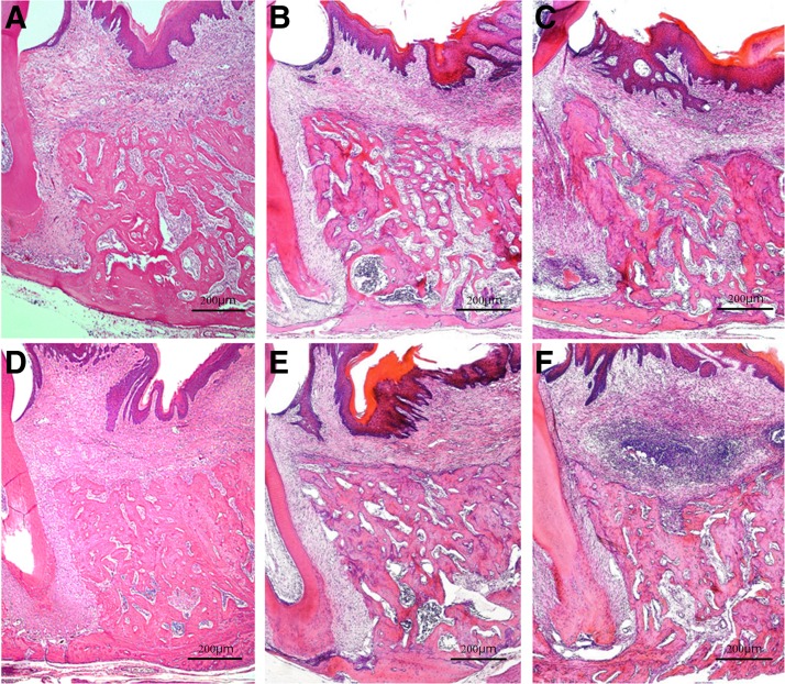
FIG. 6.
Histopathology on days 10 and 21 after tooth extraction. H&E staining; original magnification × 40. (A, D) Diode group; (B, E) CO2 group; (C, F) control group. Day 10 postextraction: extraction sockets are seen filled with newly formed bone. Numerous cells are observed in the bone marrow and the area around the cancellous bone in all groups (A–C). Day 21 postextraction: flat alveolar crest surface with almost no concavity seen in the laser treatment groups; cancellous bone is denser in the diode group than in the CO2 group (D, E). A dish-shaped concavity on the surface of the alveolar crest seen in the control group, and the less dense cancellous bone compared with that in the laser treatment groups (F).

