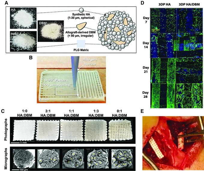
FIG. 1.
Scaffold design and implantation. (A, B) Scaffold design and fabrication. (C) Images of unimplanted scaffolds. Upper: basic geometry of the scaffolds, with 90° aligned struts and visible channels (macropores) extending through the scaffolds. Lower: cross-sections of individual struts as imaged using scanning electron microscopy (plasma-coating with ∼15 nm osmium metal, imaged with 3 kV accelerating voltage using a LEO Gemini 1525 at the EPIC facility, NUANCE Center, Northwestern University). The relative particle content [% HA (upper) vs. DBM (lower)] within the scaffolds is shown. Total particle content was held constant at 70% of the scaffold volume; the remaining 30% is PLG binder. Yellow stars identify DBM particles. (D) Laser-scanning confocal z-projections of 3D-stacks from live-dead stained scaffold seeded with primary rat bone marrow stromal cells. Blue is autofluorescence from the scaffold materials. (E) Intraoperative implantation of a scaffold. 3D, three-dimensional; DBM, demineralized bone matrix; HA, hydroxyapatite; SEM, scanning electron microscopy.

