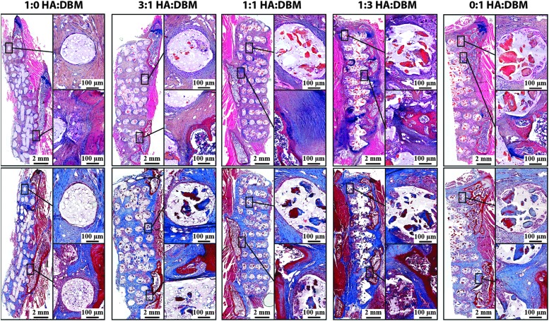
FIG. 6.
Histology. Decalcified spines were sectioned to visualize the constituents of the 8-week postoperative explanted scaffolds. The upper panel shows sections stained with alcian blue/hematoxylin/orange-eosin Y, where cartilage stains blue, bone matrix orange/red, and soft tissue stains pink. The lower panel shows sections stained with Masson's Trichrome, where collagen stains blue and bone matrix stains dark red.

