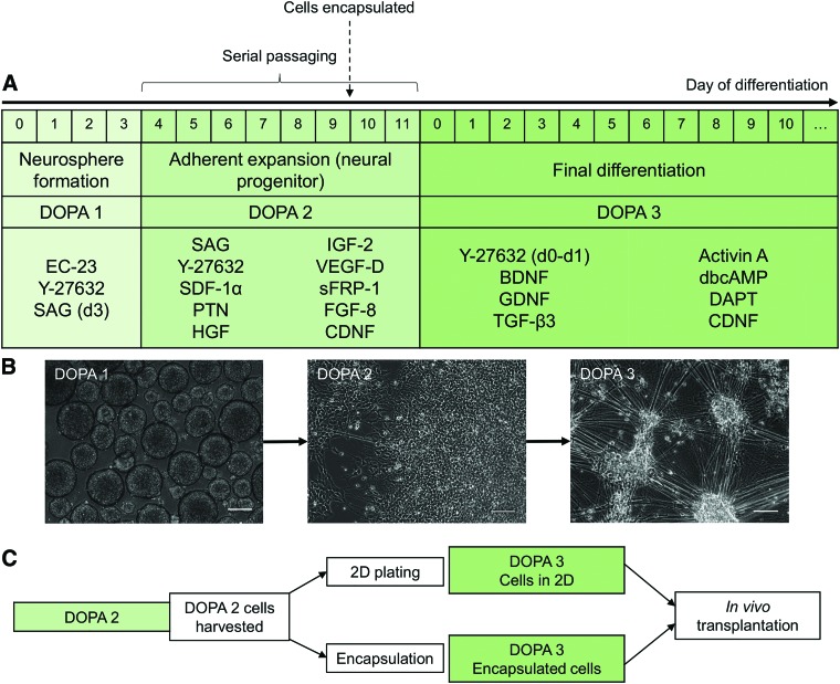
FIG. 1.
Protocol for differentiation and encapsulation of DA neurons. (A) Protocol for expansion of DA neural progenitors and final differentiation of DA neurons. (B) Representative phase-contrast images of DOPA1, DOPA2, and DOPA3 cells. Scale bar: 100 μm. (C) Timeline for encapsulation within SAPNS. DA, dopaminergic; SAPNS, self-assembling peptide nanofiber scaffolds. Color images are available online.

