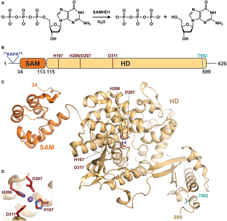
Figure 1. SAMHD1 structure and catalytic activity.
(A) The dNTP triphosphohydrolysis reaction catalysed by SAMHD1, dGTP is shown as the example substrate. (B) Domain organisation of human SAMHD1, showing the nuclear localisation signal in blue, the SAM domain in dark orange, the HD domain in light orange, the HD motif residues in maroon and phosphorylated residue Thr592 in teal. (C) Left: NMR structure of the human SAMHD1 SAM domain (PDB code: 2E8O residues 34–113). Right: X-ray crystal structure of WT human SAMHD1 HD domain (PDB code: 4BZC, monomer A, residues 115–599) [85]. The structures of the two domains are connected by a short, dotted and grey line. The HD motif-co-ordinated manganese ion is shown as a purple sphere, and the HD motif residues are shown as maroon sticks. (D) A close-up view of the HD motif-co-ordinated manganese ion in the catalytic site. PyMOL was used to prepare the structural figures.

