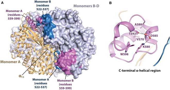
Figure 3. Thr592 phosphorylation modulates SAMHD1 tetramer stability.
(A) X-ray crystal structure of SAMHD1 HD domain tetramer (PDB code: 4BZC) [85]. Monomer A is shown in cartoon representation in light orange. Monomers B–D are shown in surface representation in grey. Residues 559–599 are in pink and residues 522–537 are in blue. (B) C-terminal, the α-helical region between residues 559–599, comprising phosphorylated residue Thr592.

