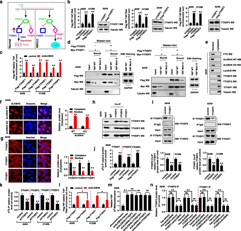
Fig. 3.
YTHDF1 and YTHDF2 competitively interacted with YTHDF3 in an m6A- independent manner to regulate YAP expression. (a) The diagram of that YTHDF1 and YTHDF2 competitively interacted with YTHDF3 to regulate YAP expression in NSCLC. (b, c) The interaction between YTHDF1/2/3 and YAP mRNA was determined by RIP assay. (d) The interactions between YTHDF1/YTHDF2 and the mRNA of YAP were increased when YTHDF3 is existed determined by RNA pulldown assay. (e) Western blot analysis indicated that FTO and ALKBH5 were only in nuclear fractions but YTHDF1/2/3 were only in cytoplasm fractions in A549 cells. (f, g) Immunofluorescent staining showed that ALKBH5 WT/KD were only in nuclear fractions (f) but YTHDF1/2/3 were only in cytoplasm fractions (g) in A549 cells. (h) Co-IPs performed using lysates collected from A549 cells with immunoprecipitation by either YTHDF1, YTHDF2 or YTHDF3 antibodies. (i) The protein level of YTHDF3 was analyzed in lysates collected from either YTHDF1 or YTHDF2 transfected A549 and H1299 cells determined by Co-IPs assays by immunoprecipitation with YTHDF2 or YTHDF1 antibodies, respectively. (j, k) The interactions between YTHDF1/2/3 and YAP mRNA were detected in A549 and H1299 cells with transfection with indicated genes determined by Co-IP assay using the MS2 coat protein system. (l) The interactions between YTHDF1/2 and YAP mRNA was determined by RIP assay in A549 and H1299 cells with transfection of indicated genes. (m) The m6A levels of YAP were analyzed by in A549 and H1299 cells with transfection into indicated genes. (n) The protein level of YTHDF3 was detected in lysates collected from A549 cells determined by Co-IP assay with immunoprecipitation with either YTHDF1 or YTHDF2 antibodies. Results were presented as mean ± SD of three independent experiments. *P < 0.05 or **P < 0.01 indicates a significant difference between the indicated groups. ns, not significant

