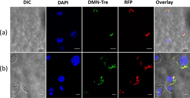
Figure 4.
Intracellular labeling of RFP-Mtb H37v with DMN-Tre. Image was taken after (a) 24 h and (b) 96 h of incubation of the Mtb-infected THP-1 cells with DMN-Tre. Images were collected in the DIC, DAPI (for NucBlue fluorescence of the nuclear stain), GFP (for DMN-Tre fluorescence), and RFP (for bacterial dTomato fluorescence) channels of the Leica SP5 laser scanning confocal microscope. Magnifications are 63× by oil immersion. Overlay represents merged images from all channels. Scale bars = 25 μm.

