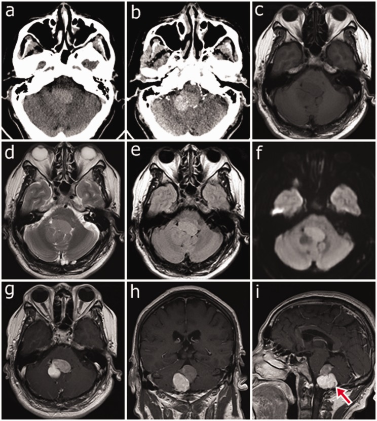
Figure 1.
(a) Unenhanced axial computed tomography showing an irregular mass in the fourth ventricle. The upper anterior part of the tumor shows slight hyperdensity, while the lower posterior part shows isodensity. (b) Contrast-enhanced axial computed tomography showing that the lower posterior part of the tumor has more pronounced enhancement than the upper anterior part. (c) Axial T1-weighted image showing an isointense mass in the fourth ventricle. (d) and (f) Axial T2-weighted image and diffusion-weighted image showing that the upper anterior part of the mass is hyperintense while the lower posterior part is hypointense, forming a black and white sign. (e) The mass is isointense on fluid-attenuated inversion recovery. (g)–(i) After gadolinium administration, the lower posterior part of the tumor has more enhancement than does the upper anterior part, forming reversed enhancement. (i) Contrast-enhanced T1-weighted sagittal image showing a flow void effect (red arrow) in the lower posterior part of the mass.

