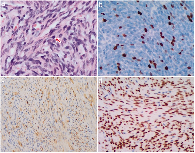
Figure 2.
Photomicrographs. (a) The tumor is composed of atypical spindle cells and a collagenized stroma, and mitotic figures can be seen (magnification = × 400, red arrow). (b) The molecular immunology borstel-1 proliferation index was 15% (magnification = × 400). (c) Neoplastic cells are strongly immunoreactive to CD34 (magnification = × 200). (d) Neoplastic cells are strongly immunoreactive to nuclear STAT6 (magnification = × 200).

