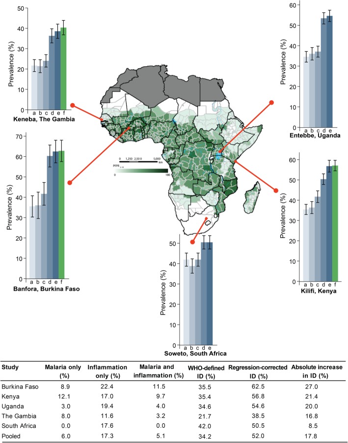
Fig. 3.
Prevalence of estimated iron deficiency across the study sites. The map shows the predicted posterior predictions of age-standardized P. falciparum prevalence (PfPR2–10) as previously published by Snow et al. [30]. Map was reproduced with permission. Graph letter “a” indicates prevalence of iron deficiency using the WHO definition, “b” excluding children with inflammation, “c” adjusting for malaria only, “d” adjusting for inflammation only, “e” adjusting for both malaria and inflammation, and “f” using transferrin saturation cut-off of < 11%. Values indicate prevalence. Malaria only indicates percentage of children with malaria parasitemia without inflammation, inflammation only as percentage with inflammation and no parasitemia, and malaria and inflammation as percentage with both parasitemia and inflammation. Absolute increase in iron deficiency was calculated as the difference between regression-corrected prevalence (corrected for both malaria and inflammation) and WHO-defined prevalence. Error bars indicate 95% confidence intervals

