
Figure 4.
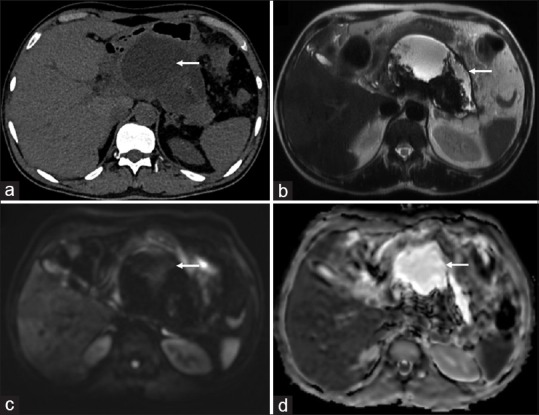
A 60-year-old male presenting with pancreatitis. Sterile collection confirmed on fine-needle aspiration. (a) Axial non-contrast CT scan showing collection (arrow) in lesser sac (b) Axial T2-weighted MRI image confirming the walled-of necrotic collection (arrow) with thick hypointense debris (c) No bright signal (arrow) is seen in the collection on diffusion-weighted images (b = 1000 s/mm2) (d) Corresponding high signal on the apparent diffusion coefficient map (arrow) is seen suggestive of no diffusion restriction