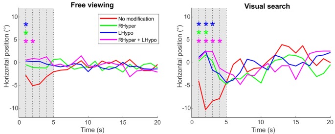
Figure 4.
Center of fixation over the trial’s time. The center of fixation is depicted as the median horizontal gaze position on the screen (y-axis) in dependence of the trial’s time (x-axis), i.e., in 1-s-bins over the 20 s of stimulus presentation duration, separately for the two task conditions and the four types of modification. Significant differences between each of the lateralized modifications (color-coded) and the NoMod baseline condition at each second of the trial’s time are marked with an * (ANOVA with post hoc t-tests, Bonferroni-corrected p < 0.05). Please note that the initial leftward bias (“pseudoneglect”) in the participants’ baseline condition (NoMod, red line), evident in the first 5 s (gray area) after the onset of the stimulus image, is counterbalanced by the three saliency modifications that favor exploration of the right hemispace.

