Figure 1.
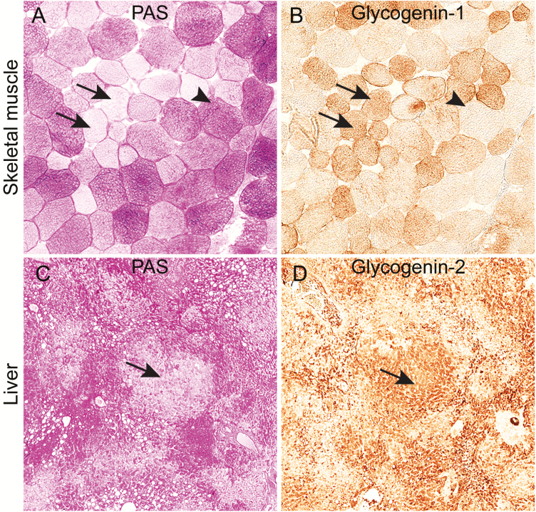
Glycogen storage and immunohistochemical expression of glycogenin in serial sections of skeletal muscle and liver of controls without glycogenin deficiency. A, Analysis of glycogen storage by staining with periodic acid–Schiff (PAS) reagent in a control muscle sample showing some pale muscle fibers (arrows) in which glycogen has been used, and also fibers with normal glycogen content (arrowhead). B, Immunohistochemical staining using an antibody to glycogenin-1. In the fibers depleted of glycogen, the glycogenin-1 molecules, which are normally covered by polysaccharide chains, are exposed and expression of glycogenin-1 is detectable (arrows). Fibers with normal glycogen content do not show glycogenin-1 expression by immunohistochemical staining (arrowhead). C, PAS staining of control liver showing pale regions (arrow) where glycogen has been used and the glucose molecules are digested. D, Immunohistochemical staining using an antibody to glycogenin-2. In the pale regions in the PAS staining, glycogenin is exposed and the expected expression of glycogenin-2 is evident (arrow).

