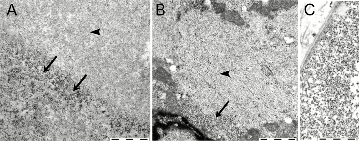
Figure 6.
Electron microscopy of glycogen storage in cardiac and skeletal muscle in patients with biallelic GYG1 mutations. A, In skeletal muscle, the accumulated material consisted of a mixture of granular, apparently normal glycogen (arrows) and fibrillar material (arrowhead). The filamentous material represented large regions of the periodic acid–Schiff–positive storage material and was morphologically compatible with polyglucosan. B, In cardiac muscle, there was a similar mixture of granular material (arrow) and fibrillar material (arrowhead) material. C, Normal muscle glycogen is shown for comparison. Bars correspond to 1 µm.

