Fig 2.

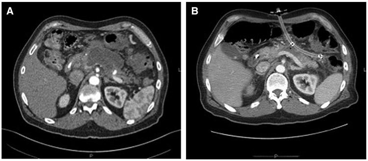
Radiographic representation of U-tube drainage. A, Initial necrosis prior to drain placement. B, Placement of U-tube with resolution of necrosis and large-bore fistulous tract visible anteriorly and laterally.

Radiographic representation of U-tube drainage. A, Initial necrosis prior to drain placement. B, Placement of U-tube with resolution of necrosis and large-bore fistulous tract visible anteriorly and laterally.