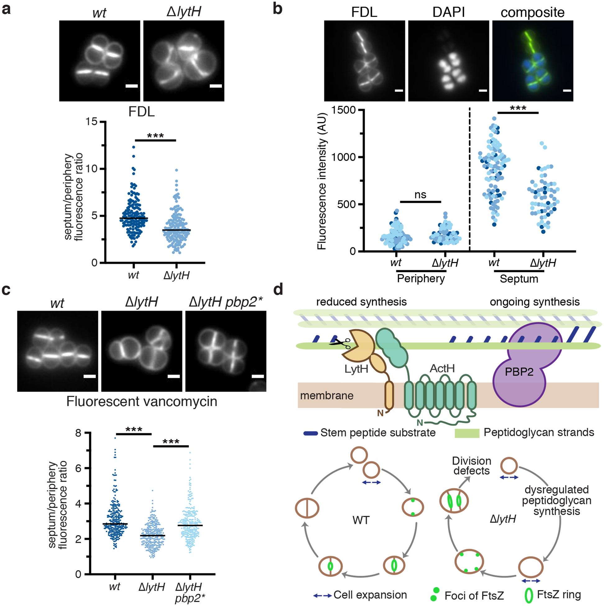
Figure 4: LytH spatially regulates peptidoglycan synthesis.

a, Cells were labeled with fluorescein-D-lysine (FDL) to mark sites of transpeptidase activity; ΔlytH cells showed decreased septal to peripheral signal. Fluorescence ratios were calculated using only cells with a single complete septum. FDL incorporation was catalyzed by the transpeptidases PBP1, PBP2, and PBP3; pbp4 was deleted to reduce background47. In all plots, the median is marked by a black line, each dot represents a single cell, and p-values were determined by two-sided Mann-Whitney U tests. In this plot: n = 159 (wt) and 167 (ΔlytH) cells; P = 1.34×10⁻¹² (***P < 0.001). Scale bars, 1 μm. b, A mixed population of WT and ΔlytH cells was labeled with FDL and imaged in the same frame to directly compare the signal at the septum and periphery; ΔlytH cells showed decreased septal activity. In these images, ΔlytH cells were stained with DAPI to distinguish them from WT cells. Fluorescence intensity measurements correspond to the median intensity at the periphery or septum. Different colored dots represent cells analyzed from different fields of view: n = 100 (wt) and 61 (ΔlytH) cells. From left to right: P = 0.0864 and 4.49×10⁻¹⁰ (***P < 0.001; ns, not significant). Scale bars, 1 μm. c, Newly synthesized peptidoglycan was labeled with fluorescent vancomycin; a decrease in septal to peripheral signal was observed for ΔlytH cells expressing pbp2WT. pbp2* represents the suppressor allele pbp2F158L. From left to right: P = 3.87×10⁻³² and 1.51×10⁻¹⁹ (***P < 0.001); counted 283 (wt), 287 (ΔlytH), and 278 (ΔlytH pbp2*) cells. Scale bars, 1 μm. d, Model for LytH function. Control of cell size is required for FtsZ localization at midcell to properly initiate division. Free stem peptides limit rapid diffusion of PBP2 to midcell during division, leading to increased cell size due to dysregulated peptidoglycan synthesis. LytH regulates stem peptide abundance on membrane-proximal peptidoglycan: in its absence, cells enlarge, FtsZ becomes mislocalized, and division initiates at aberrant sites. Data are representative of two (a-c) independent experiments.
