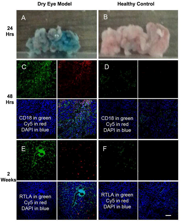
Figure 1.

Biodistribution of subconjunctivally injected D-Cy5 in LG. Co-localization of the Cy5 signal (red) with inflammatory cell marker staining (green) as CD 18 (A, D, G) and RTLA (B, E, H) were found in LG with established disease. The upper panel (A-C), the middle panel (D-F) and the lower panel (G-I) represent samples from 24 hours, 72 hrs, and 2 weeks post-subconjunctival injection, respectively. Minimal uptake was found in LG of normal control group (C, F, I). Blue for DAPI staining. Scale Bar :100 µm.
