
Fig 2. Measles infection enhances vomocytosis of C. neoformans.
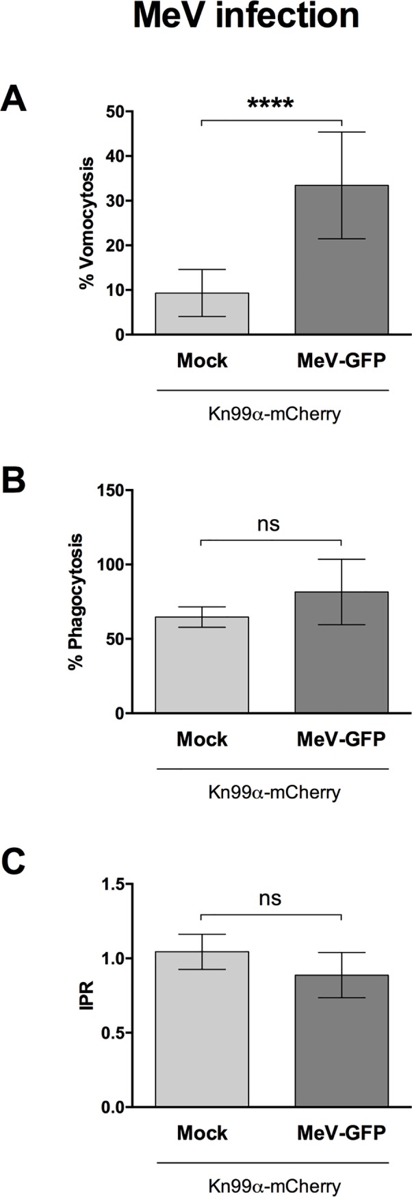
Human monocyte-derived macrophages were infected with Measles virus and subsequently infected with C. neoformans. Time-lapse microscopy videos were manually scored for vomocytosis (top), uptake (middle) and intracellular proliferation rate of C. neoformans (bottom). A Graph shows percentage of Cryptococcus-infected macrophages which have experienced at least one vomocytosis event. B Percentage of Cryptococcus-infected macrophages. C Intracellular proliferation rate of C. neoformans over 18 hours. In all cases, pooled data from 3 independent experiments is shown. Categorical vomocytosis and phagocytosis data was analysed by Chi2 test followed by Fisher's exact test. **** p < 0.0001. IPR data was analysed using Mann-Whitney test.