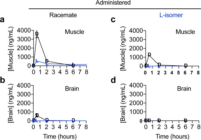
Fig 6. Graphs of the concentration of enantiomers in tissue versus time after administration of racemic N-acetyl-DL-leucine or purified N-acetyl-L-leucine.
Data are for (a,b) muscle and (c,d) brain and presented as linear-linear plots. Values are the mean ± standard error of the mean with n = 3 (mice).

