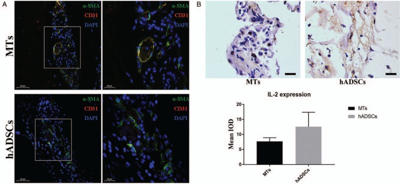
Figure 5.

Immunofluorescent double staining of α-SMA and CD31 for the detection of neo-vascularization. (A) After 7 days of subcutaneous implantation, CD31+ cells were observed localized in and around the α-SMA-positive vascular-like structure. Higher magnification of the boxed area in left panel further demonstrated the location of the CD31+ cells (right). Scale bar = 50 and 20 μm. (B) Immunohistochemistry staining of IL-2 revealed that the retrieved structure constructed from MTs tended to express IL-2 at lower levels than their counterpart made from hADSCs (top). Semi-quantitative analysis indicated that there was no statistical significance in the difference of IL-2 expression between these two groups (bottom). Scale bar = 20 μm. α-SMA: α-Smooth muscle actin; IL-2: Interleukin-2; IOD: Integral optical density; MTs: Microtissues; hADSCs: Human adipose-derived stem cells.
