
Figure 8.
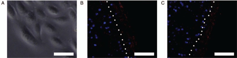
Morphology of urothelial cells and immunofluorescent staining of cytokeratins AE1/AE3 for encapsulated structures containing non-induced MTs/induced MTs seeded with urothelial cells. (A) Inverted microscopy demonstrated the cobblestone appearance of the human UCs in culture. (B) Immunofluorescent staining of UCs seeded onto encapsulated structures containing non-induced MTs were positive for cytokeratins AE1/AE3 (red). (C) Immunofluorescent staining of UCs seeded onto encapsulated structures containing non-induced MTs were positive for cytokeratins AE1/AE3 (red). After 1 week of seeding, UCs formed mainly a monolayer on the surface of the encapsulated structures (right of the white dotted line). Scale bar = 50 μm. MTs: Microtissues; UCs: Urothelial cells.