
Figure 3.
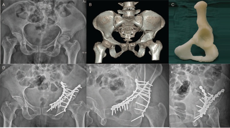
Images of one case in the 3D printing group. (A) Pre-operative radiograph (anteroposterior view). (B) The 3D reconstructed computed tomography images. (C) The 3D printed model used for pre-operative evaluation. (D–F) Post-operative follow-up radiograms at 1 year (pelvic anteroposterior and Judet views). 3D: Three-dimensional.