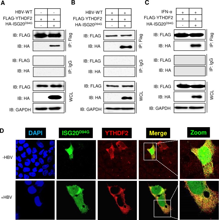
Fig 2. HBV-independent interaction between YTHDF2 proteins with ISG20.
(A) FLAG-YTHDF2 and HA-ISG20D94G plasmids were co-transfected into HepG2 cells. After 48h of incubation cells were harvested and lysates were prepared. FLAG antibody was used for IP and then probed with FLAG and HA antibody after Western Blot. (B) FLAG-YTHDF2 and HA-ISG20D94G plasmids were co-transfected into HBV expressing HepG2 cells. After 48 hour of incubation cells were harvested and lysates were prepared. IP was done with FLAG antibody and then probed with FLAG and HA antibody after Western Blot. (C) FLAG-YTHDF2 and HA-ISG20D94G plasmids were co-transfected into HepG2 cells and then incubated for 48h until harvest and IFN-α was added (2000 IU/ml) 24h before harvesting the cells. After preparing the lysates IP was done with FLAG antibody and probed with FLAG and HA antibody after Western Blot. (D) Confocal microscopy of HepG2 cells transfected with HA-ISG20D94G and FLAG-YTHDF2, showing signals for DAPI stained nuclei (blue), ISG20D94G (green), YTHDF2 (red) and the merged images (yellow) (upper panel). Confocal microscopy of HBV expressing HepG2 cells transfected with HA-ISG20D94G and FLAG-YTHDF2, showing signals for DAPI stained nuclei (blue), ISG20D94G (green), YTHDF2 (red) and the merged images (yellow) (lower panel). The data for this figure are from two independent experiments and the bars represent the mean ± SD.

