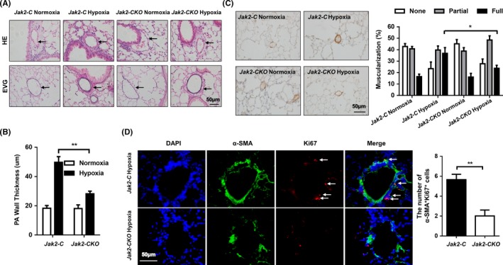
Figure 4.

Loss of Jak2 in smooth muscle cells protected against pulmonary vascular remodelling after hypoxia. Representative HE‐stained (top) and EVG‐stained (bottom) sections (A), quantification of pulmonary arteriole wall thickness (B) and α‐SMA immunostaining (C) in the lungs of Jak2‐C and Jak2‐CKO mice after normoxic (n = 8 per group) or hypoxic (n = 10 per group) exposure for 28 days. Ten vessels were analysed per mouse. Coimmunostaining results of α‐SMA and Ki67 in lung sections from Jak2‐C and Jak2‐CKO mice after hypoxic (D; n = 10 per group) exposure for 28 days. All images were taken at an original magnification of ×400. The data are represented as the mean ± SEM. *P < .05; **P < .01. HE, haematoxylin and eosin; EVG, elastic van gieson; PA, pulmonary artery
