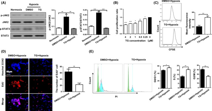
Figure 5.

Hypoxia‐induced HPASMC proliferation was suppressed by a JAK2 inhibitor. Western blot analysis of p‐JAK2, JAK2, p‐STAT3 and STAT3 in HPASMCs (A). CCK‐8 analysis of HPASMCs pre‐treated with different concentrations of TG for 1 h following 24 h hypoxic exposure (B). CFSE dilution analysis (C), EdU staining (D) and cell cycle analysis (E) of HPASMCs pre‐treated with DMSO or TG for 1 h following 24 h hypoxic exposure. All images were taken at an original magnification of ×400. The data are represented as the mean ± SEM. *P < .05; **P < .01; ***P < .001. TG, TG‐101348; CFSE, carboxyfluorescein diacetate succinimidyl ester; EdU, 5‐ethynyl‐uridine; DMSO, dimethyl sulphoxide
