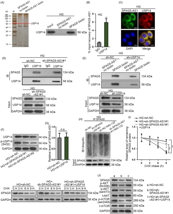
Figure 6.

SPAG5‐AS1 stabilized SPAG5 protein through interacting with USP14 in HG‐treated HPCs. A, The silver staining gel manifested that a protein band in the pull‐down products of SPAG5‐AS1 biotin group was observed in HG‐treated HPCs. Western blot assay for the USP14 enrichment in the pulldown of SPAG5‐AS1 biotin group and SPAG5‐AS1 no‐biotin group in HG‐treated HPCs. B, RIP analysis confirmed the SPAG5‐AS1 level in USP14‐binding complexes. C, FISH and IF staining were performed for the localization of SPAG5‐AS1 and USP14 protein in HG‐treated HPCs. Scale bar: 10 μm. D, Co‐IP assay of the SPAG5 and USP14 enrichments in the precipitated products of anti‐USP14, under SPAG5‐AS1 depletion or sh‐NC control. E, Western blot for the SPAG5 and USP14 abundance in the pulldown of SPAG5‐AS1 biotin group and no‐biotin group under USP14 knockdown or sh‐NC control. F, Silence of SPAG5‐AS1 reduced the level of p‐USP14 (S432) without changing its total protein level in HG‐treated HPCs. G, Knockdown of USP14 failed to impact SPAG5‐AS1 level in HG‐treated HPCs. H, The immunoblot of ubiquitin in the precipitates of SPAG5 and the input level of SPAG5 under the silence of SPAG5‐AS1 in HG‐treated HPCs treated with or without MG‐132. I, Remaining SPAG5 protein level after the treatment of CHX was detected by Western blot and quantitated at 0, 3, 6 and 9 h in HG‐treated HPCs transfected with sh‐NC, sh‐SPAG5‐AS1#1 or sh‐SPAG5‐AS1#1 + pcDNA3.1/USP14. J, The protein level of SPAG5 as well as phosphorylated and total levels of AKT and mTOR in HPCs treated with HG + sh‐NC, HG + sh‐SPAG5‐AS1#1 or HG + sh‐SPAG5‐AS1#1 + USP14. All experiments were conducted in triplicates. Data are presented as mean ± SD. **P < .01
