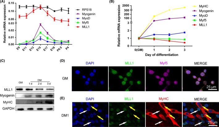
Figure 1.

Expression pattern of MLL1 during muscle development. A, qPCR measurement of mRNA expression of MLL1 and myogenic markers (Myf5, MyoD and Myogenin) in dorsal muscle of mouse at several developmental times. GAPDH was used as an internal control for normalization, and ribosomal protein S18 (RPS18) was used as a negative control. E: days of embryo age, P: days of age post‐birth. B, The mRNA levels of MLL1 and myogenic markers during C2C12 cell differentiation at several indicated time points. When the cells were cultured in growth medium (GM) at sub‐confluent densities, it was defined as day 0; when the cells reached 100% confluence, GM was changed to differentiation medium (DM). C, Western blot analysis of MLL1 protein expression was performed in C2C12 cells grown in GM, as well as 1d, 2d and 3d in DM. Western blot for the myogenic markers myogenin and MyHC was performed to ensure that myogenic differentiation occurred properly. GAPDH was used as a loading control. D, Immunofluorescence staining of MLL1 and Myf5 (a marker of proliferating myoblasts) in C2C12 cells cultured in GM. The cell nucleus was stained with DAPI. Scale bar = 25 μm. E, Immunofluorescence staining of MLL1 and MyHC (a marker of differentiated myotubes) in C2C12 cells differentiated for 1 day in DM. White arrows indicate nucleus of myoblasts, and yellow arrows indicate nucleus in MyHC+ myotubes. Scale bar = 25 μm. Data are presented as mean ± SEM, n = 3 per group
