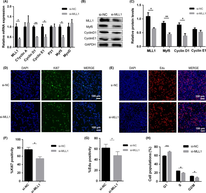
Figure 2.

MLL1 regulates myoblast proliferation through Cyclin D1. C2C12 cells transfected with negative control siRNAs (si‐NC) or MLL1 siRNAs (si‐MLL1) were cultured in GM for 2 d. A, qPCR analysed the mRNA levels of MLL1, cell cycle regulators (Cyclin A/D1/E1, P21 and DHFR) and two myogenic transcription factors (MyoD and Myf5). B, Western blot detected the protein levels of MLL1, Myf5, Cyclin D1 and Cyclin E1 in proliferating C2C12 cells treated as above. C, The relative protein levels of target proteins normalized to GAPDH signals in (B) were obtained through Western blot (WB) band grey scanning. D, Immunofluorescent staining of Ki67 (a marker of proliferation) was performed to examine proliferation ability of si‐MLL1 C2C12 cells. Scale bar = 100 μm. E, Representative images of the EdU staining for si‐MLL1 C2C12 cells. Scale bar = 100 μm. F‐G, The percentages of EdU‐ and Ki67‐positive cells compared with the total number of nuclei were presented, respectively. H, The cell cycle phase of si‐NC and si‐MLL1 C2C12 cells was examined through flow cytometry analysis after PI staining. Data are presented as mean ± s.e.m., n = 3; *P < .05, **P < .01 (Student's t test)
