Figure 4.

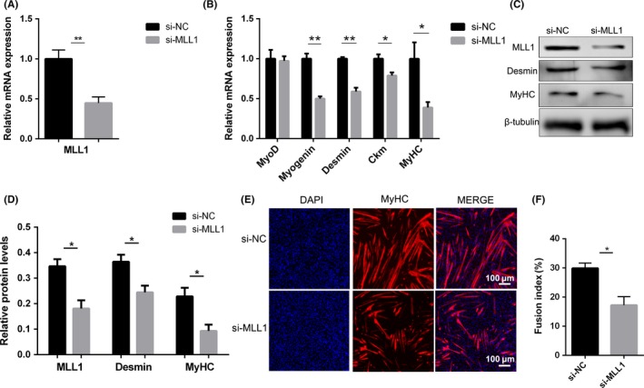
Knockdown of MLL1 impairs myogenic differentiation. C2C12 cells were transfected with si‐NC or si‐MLL1, and then induced to differentiate in DM for 3 days. A‐B, qPCR was performed to detect the mRNA levels of MLL1 as well as myogenic differentiation markers. C, Western blot detected the protein levels of MLL1, Myogenin and MyHC. β‐tubulin was used as a loading control. D, The relative protein levels of target proteins normalized to β‐tubulin signals in (C) were obtained through WB band grey scanning. E, Immunofluorescent staining for MyHC in si‐NC or si‐MLL1 C2C12 cells was performed to detect myotube formation. The cell nucleus was stained with DAPI. Scale bar = 100 μm. F, The fusion index (the percentage of nuclei in fused myotubes out of the total nuclei) in (E) was calculated. For each group, six random microscopic fields were selected randomly. Data are showed as mean ± SEM, n = 6 per group. *P < .05, **P < .01 (Student's t test)
