Figure 1.

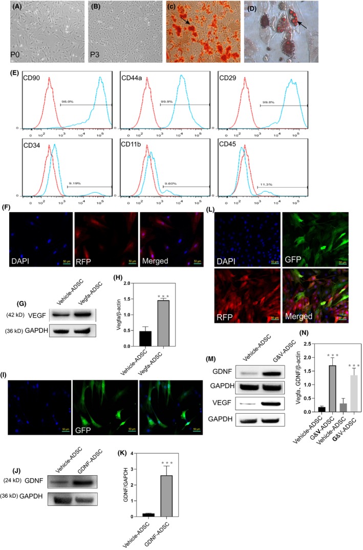
Genetically modified ADSCs produced more VEGF and GDNF. A,B, Morphology of ADSCs in passage 0 and passage 3. C,D, Adipose‐derived stem cells exhibited pluripotency after induction, as evidenced by the presence of typical phenotype of osteocytes (stained with Alizarin Red S) and the typical phenotype of adipocytes (stained with Oil Red O). E, Flow cytometry showed the ADSCs expressed more stem cell markers (CD90, CD44a and CD29), but less haematopoietic and endothelial markers (CD34, CD11b and CD45). F, Immunofluorescence analysis of the expression of RFP in transfected ADSCs. G,H, Western blot analysis showed markedly increased the levels of VEGF in transfected ADSCs. I, Immunofluorescence analysis of the expression of GFP in transfected ADSCs. J,K, Western blot analysis showed increased the levels of GDNF in transfected ADSCs. L, Immunofluorescence of ADSCs that co‐expressed RFP and GFP. M,N, Western blot analysis of ADSCs co‐transfected with VEGF and GDNF. All values are represented as the mean ± SD from three independent experiments, each with three replicates. Statistically significant differences from the control group are denoted as follows: ***P < .001, **P < .01 and *P < .05 (independent samples t test)
