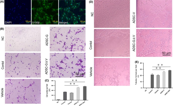
Figure 2.

Adipocyte‐derived stem cells transfected with VEGF and GDNF enhanced tubule formation of HUVECs and chemotaxis of primary SCs, respectively. A, Isolation and identification of primary SCs from rat sciatic nerves (passage 2). B, Schwann cell chemotaxis in response to different cell culture supernatants by crystal violet staining and visualization using an inverted microscope. C, Quantification of migrated cells. D, Tubule formation of HUVECs cultured with different supernatants. E, Quantitative analysis of tubular formation of HUVECs. Each bar depicts the mean ± SD (***P < .001, **P < .01 and *P < .05, n = 3); one‐way ANOVA followed by the S‐N‐K test
