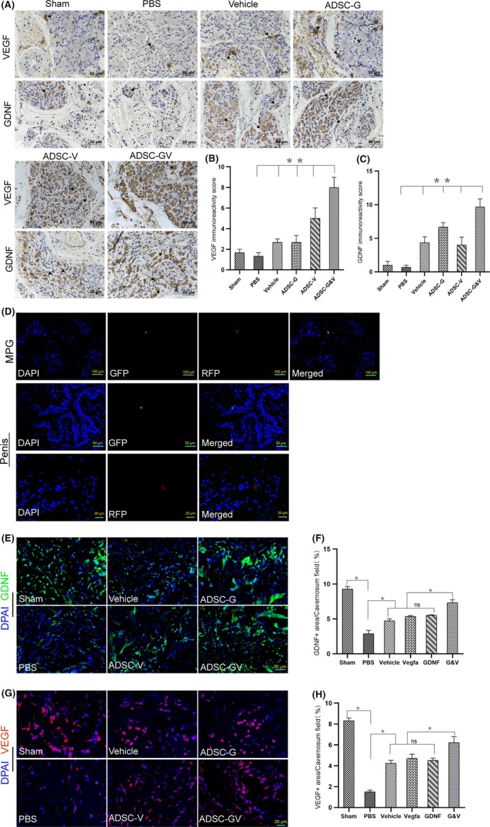
Figure 3.

The expression of VEGF and GDNF in the MPG and penis after transplantation of GM‐ADSCs. A, Immunohistochemical staining of rat MPG tissue cross‐sections from each animal in the respective groups using specific VEGF and GDNF antibodies. Arrows indicate typical immune‐positive cells. B,C, Immunohistochemical score was obtained by analysing the staining intensity and positive rate of VEGF and GDNF. D, After 14 d, transfected ADSCs were visible in the MPG and corpus cavernosa. E, Representative immunofluorescence staining of GDNF (green) in a penile mid‐shaft specimen 2 wk after BCNI and treatment. F, Quantitative analysis of the GDNF‐positive area. G,H, Representative immunofluorescence of VEGF in a penile mid‐shaft specimen, and quantitative analysis of the VEGF immunofluorescence‐positive area. Data are depicted as the mean ± SD from n = 6 animals per group (*P < .05). Immunofluorescence was analysed using one‐way ANOVA followed by the S‐N‐K test. Quantitative analysis of immunohistochemistry was performed using the Kruskal‐Wallis H test
