
Fig. 1.
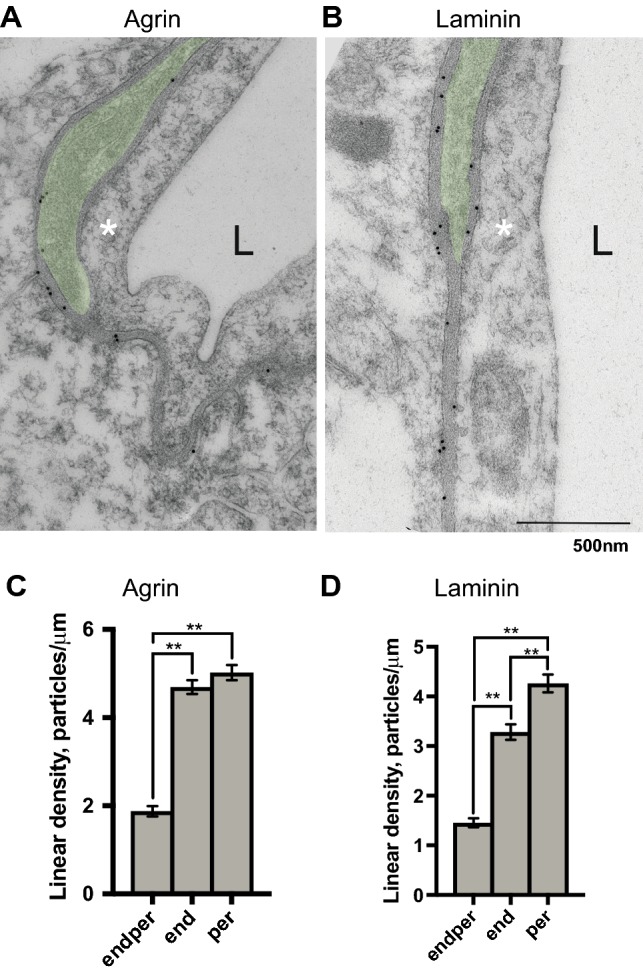
Evidence of heterogeneous labelling intensity of basal lamina microdomains around capillary bed for agrin and laminin is shown. a, b Representative electron micrographic images of anti-agrin and anti-laminin immunogold labelling around capillary bed. Endothelium (annotated with asterisk), capillary lumen (L) and part of a pericyte (coloured green), surrounded by a basal lamina on both sides are shown. Astrocyte endfeet abut the basal lamina overlying both pericytes and endothelium. Immunogold particles directed against agrin (a), were quantified as gold particles per unit length (μm) for all of the depicted basal lamina microdomains (c); between astrocytes and endothelium (end mean = 4.692, Std error = 0.158), between astrocytes and pericytes (per mean = 5.022, Std error = 0.172) and between pericytes and endothelium (endper mean = 1.878, Std error = 0.117). Labelling intensity differs depending on the adjoining cell type and is notably more intense in the two basal lamina microdomains abutting astrocytes compared with that interlaced between pericytes and endothelium. This difference (linear density, graph c) is statistically significant (p < 0.001). No difference is found between the end and per microdomains. Immunogold labelling against laminin (b, d) displays a similar pattern (end mean = 3.282, Std error = 0.158; per mean = 4.263, Std error = 0.181; endper mean = 1.456, Std error = 0.092). Differences between all three basal lamina microdomains are statistically significant (graph d, p < 0.001) and labelling is most pronounced in the basal lamina interlaced between astrocyte endfeet and pericytes. Scalebar 500 nm. **Signifies p < 0.001. Error bars represent standard error of the mean (SEM)