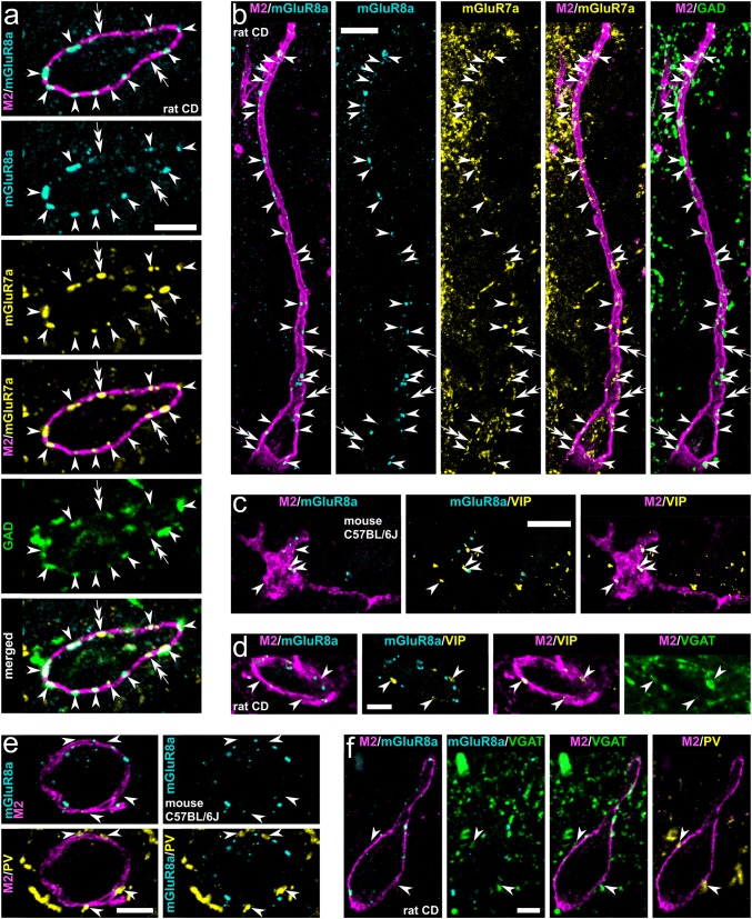
Fig. 2.
Molecular characterisation of inputs to hippocampal trilaminar neurons in rat (a, b, d, f) and mouse (c, e). a, b Group III metabotropic glutamate receptors mGluR7a and mGluR8a are co-expressed in the majority of GABAergic GAD+ boutons (arrowheads) targeting trilaminar cells in the CA3 and CA1 (maximum intensity projections, z stacks, heights 0.7 μm and 3.5 μm, respectively). Note also some mGluR7a+/GAD−/mGluR8a− input terminals (double arrows). c, d Trilaminar cells are innervated by mGluR8a− GABAergic terminals (arrowheads) co-expressing VIP and VGAT (maximum intensity projections, z stacks, heights 1.7 μm). e, f Trilaminar cells are innervated by mGluR8a− GABAergic terminals (arrowheads) co-expressing PV and VGAT (maximum intensity projection, z stack, height 2.3 μm; and confocal microscopic single optical section, 0.3 μm). CD, Sprague–Dawley; +, immunopositive; −, immunonegative; scale bars 5 μm in a, d–f 10 μm in b, c

