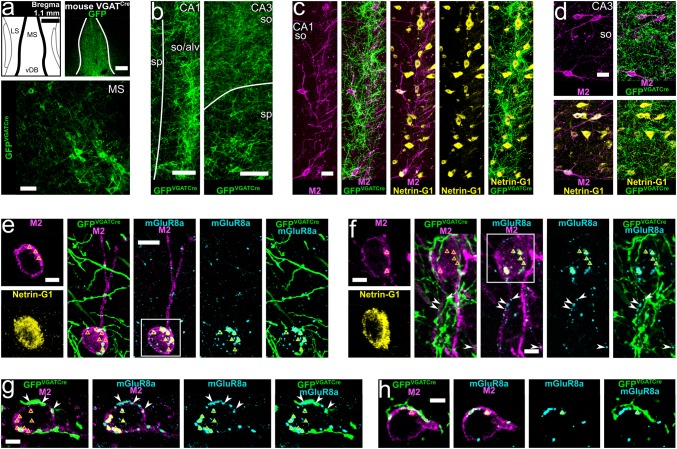
Fig. 4.
GABAergic neurons of the medial septum innervate hippocampal trilaminar cells. a Viral injection site and expression of GFP (top right, single optical section, 20.8 μm) together with labelled GABAergic neurons in the medial septum of a VGATCre mouse (GFPVGATCre+ ; bottom, maximum intensity projection, z stack, height 13 μm). b–d GFPVGATCre+ medial septal axons in areas CA1 and CA3 in apposition to cell bodies and dendrites of M2+/Netrin-G1+ neurons (maximum intensity projections, z stacks, heights 35.9 μm, 16.2 μm and 27.2 μm). e–h M2+/Netrin-G1+ neurons postsynaptic to GFPVGATCre+ medial septal boutons innervated by mGluR8a+ terminals identifying them as trilaminar cells (maximum intensity projections, z stacks, heights 10.8 μm, 5.2 μm, 5.4 μm and 4.9 μm). Note mGluR8a expression in some of the GFPVGATCre+ medial septal input terminals (arrowheads). In most neuronal somata we detected high levels of lipofuscin (triangles). MS, medial septum; LS, lateral septum; vDB, vertical diagonal band; so, stratum oriens; alv, alveus; sp, stratum pyramidale;+ , immunopositive; scale bars 250 μm in top of a 50 μm in bottom of a 100 μm in b, 25 μm in c, d, 10 μm in e, 5 μm in f–h, and insets of e, f

