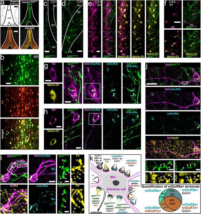
Fig. 5.
Medial septal neurons expressing PV innervate trilaminar cells in the hippocampus. a Viral injection site and expression of GFP together with PV immunoreactivity in the medial septum of a PVCre mouse (single optical section, 20.8 μm). b Co-expression of PV and GFP in neurons of the medial septum (GFPPVCre+ ; maximum intensity projection, z stack, height 19.8 μm). c–f Axonal projections of GFPPVCre+ medial septal neurons in areas CA1 and CA3 in apposition to cell bodies and dendrites of M2+/Netrin-G1+ neurons (maximum intensity projections, z stacks, heights 31.2 μm, 31.2 μm, 11.9 μm and 6.8 μm). g–i M2+/Netrin-G1+ neurons postsynaptic to GFPPVCre+ medial septal terminals (arrowheads) innervated by mGluR8a+ boutons identifying them as trilaminar cells (maximum intensity projections, z stacks, heights 4.6 μm, 4.2 μm and 9.2 μm). Expression of PV confirmed in the GFPPVCre+ medial septal terminals (i). In most neuronal somata we detected high levels of lipofuscin (triangles). j Expression of VGAT in the GFPPVCre+ medial septal terminals (arrowheads) targeting hippocampal trilaminar cells (maximum intensity projection, z stack, height 5.3 μm). k Summary schematic diagram of the identified synaptic inputs innervating M2+ trilaminar cells. Right: quantification of mGluR7a and GAD in mGluR8a+ terminals targeting trilaminar cells. MS, medial septum; LS, lateral septum; vDB, vertical diagonal band; hDB, horizontal diagonal band; so, stratum oriens; sp, stratum pyramidale; sl, stratum lucidum; sr, stratum radiatum; + , immunopositive; scale bars, 250 μm in a, 50 μm in b–d, f, 25 μm in e, j, 10 μm in g–i and insets of g, h, 2.5 μm in insets of i, j

