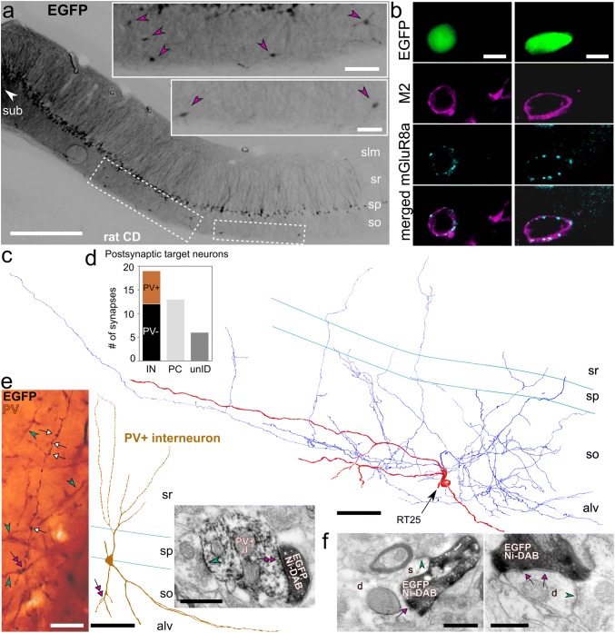
Fig. 6.
Retrogradely labelled hippocampo-subicular projecting trilaminar neurons in stratum oriens of the rat CA1 area. a Neurons in the CA1 area expressing EGFP following the injection of a recombinant adenovirus vector into subiculum (left, white arrowhead). Note some putative GABAergic neurons (insets, magenta arrowheads) retrogradely labelled together with the majority of CA1 pyramidal cells (contrast inverted epifluorescence images). b Hippocampo-subicular projecting EGFP-labelled cells in stratum oriens co-expressing M2 and decorated by mGluR8a+ terminals (epifluorescence micrograph). c Single-section reconstruction of soma with dendrites (red, section thickness 70 μm) and local axon collaterals (blue) of one EGFP-labelled GABAergic neuron co-expressing M2 and innervated by mGluR8a+ terminals (b left). d Postsynaptic target distribution of the EGFP-labelled cell reconstructed in c. eleft Axon collateral with boutons (white arrows) of the EGFP-labelled cell (black, HRP with nickel-DAB) reconstructed in c and PV+ dendrites (cyan arrowheads, brown, HRP with nickel-free DAB) in stratum oriens (transmitted light micrograph). Note the contact (magenta double arrow) between one EGFP-labelled bouton and one dendrite of a local PV+ interneuron (middle, reconstruction of soma and dendrites). Right: electron micrograph of the same EGFP-labelled bouton (black, HRP with nickel-DAB) making a type II synapse (magenta double arrow) with the PV+ interneuron dendrite (HRP with nickel-free DAB) also receiving a type I synapse (cyan arrowhead) of unknown origin. f Electron micrographs of EGFP-labelled boutons (black, visualised using HRP reaction and nickel-DAB as chromogen) of the cell reconstructed in c making type II synapses (magenta arrows) with a pyramidal cell dendrite (left, d) and an interneuron dendrite (right, d). Note the type I synapses (cyan arrowheads) received onto the spine (left, s) emanating from the dendrite and onto the shaft (right), respectively, identifying the postsynaptic targets as pyramidal cell and interneuron. CD, Sprague–Dawley; IN, interneuron; PC, pyramidal cell; unID, unidentified; + , immunopositive; −, immunonegative; slm, stratum lacunosum-moleculare; sr, stratum radiatum; sp, stratum pyramidale; so, stratum oriens; alv, alveus; sub, subiculum; scale bars 0.5 mm in a, 100 μm in insets of a and for reconstructions in c, e, 10 μm in b and for epifluorescence images in c, 0.5 μm in d and for electron micrograph in e, 25 μm for light micrograph in e

