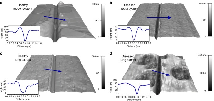
Fig. 3. Film thickness of model systems and lung extracts.
AFM 3D height images and cross-sectional profiles of a healthy model systems composed of 3:1 DPPC:DOPG, scan size μm × μm, b diseased model system consisting of 3:1 DPPC:DOPG with addition of cardiolipin and Ca, scan size μm × μm, c healthy bovine lipid–protein extract surfactant (BLES), scan size μm x μm, and d diseased BLES with cardiolipin and Ca. Blue arrows display scanning directions of profiles, scan size μm × μm.

